CBM Dibangou, RJM Ezabwanga, AD Ndiaye, NM Sambou
09-Nov-2026
Study design: Case report and review of the literature. Objective: To report a rare location of penetrating traumatic spinal injury and to review the literature. Background: Apart from gunshot wounds, penetrating spinal injuries are rare, and even rarer at the level of the sacrum. Few cases have been reported in the literature, and their management has not been standardised. Case presentation: A 22-year-old man was struck in the lower back with a screwdriver during a fight. He presented to the emergency department with part of the screwdriver still in place and was neurologically intact. A plain radiograph showed the radiopaque shaft embedded in the body of S3. After skin preparation, the screwdriver was removed under local anaesthesia and the entry wound was sutured. The patient received a tetanus booster and intravenous antibiotic prophylaxis. Outcome: The patient was discharged after 24 hours of uneventful observation, and his neurological examination remained normal throughout follow-up. Conclusion: Given the anatomy of the sacrum and our review of the literature, removal of a foreign body retained in the spine without surgical exploration may be considered in the absence of neurological symptoms, cerebrospinal fluid (CSF) leakage, or a foreign body completely buried beneath the skin.
Penetrating Trauma, Stab Wound, Sacrum, Case Report
Type d'étude : Rapport de cas et revue de la littérature.
Objectif : Présenter une localisation rare de lésion traumatique pénétrante du rachis, ainsi qu'une revue de la littérature.
Contexte : Les traumatismes pénétrants du rachis sont rares en dehors des lésions par arme à feu, et plus encore au niveau du sacrum. Peu de cas sont retrouvés dans la littérature, et leur prise en charge n'est pas codifiée.
Observation : Un homme de 22 ans a reçu, au cours d'une rixe, un coup porté dans le bas du dos à l'aide d'un tournevis. Il s'est présenté aux urgences avec une partie du tournevis en place et était neurologiquement indemne. Une radiographie standard a montré la présence de la tige radio-opaque enfouie dans le corps de S3. Après préparation cutanée, le tournevis a été extrait sous anesthésie locale et l'orifice d'entrée a été suturé. Le patient a reçu un rappel de vaccin antitétanique et une antibioprophylaxie par voie intraveineuse.
Résultat : Le patient a été libéré après une surveillance de 24 h sans complication, avec un examen neurologique resté normal au cours du suivi.
Conclusion : Au regard de l'anatomie du sacrum et de notre revue de la littérature, il convient de considérer le retrait du corps étranger retenu dans le rachis sans exploration chirurgicale en l'absence de symptomatologie neurologique, de fuite de liquide cérébrospinal (LCS) ou de corps étranger enfoui complètement sous la peau.
Mots-clés : Traumatisme pénétrant ; plaie par arme blanche ; sacrum ; rapport de cas.
An injury is described as penetrating when the wounding agent passes through the skin to reach the underlying anatomical structures [6].
Penetrating spinal injuries are uncommon in most regions, except in South Africa, where Peacock et al. published a series of 450 cases collected over 13 years [18]. The most frequent cause of penetrating spinal injury reported in the literature is gunshot (ballistic) injury [5, 15]. This is favoured by the accessibility of firearms, particularly in developed countries, whereas non-ballistic penetrating injuries are less frequent [4]. To our knowledge, the only series on this subject in sub-Saharan Africa is the study by Gueye et al. in 1998, which reported 6 cases of penetrating spinal injury in a series of 16 patients [8].
The most frequent locations are cervical and thoracic [18, 24].
We report a case of penetrating spinal injury at an unusual location and discuss the parameters to be assessed for rapid and effective management.
Our patient, a 22-year-old man with no significant medical history, presented to the emergency department of Ziguinchor Regional Hospital at 10 a.m., following an altercation with a friend that reportedly took place the previous evening at around 11 p.m. The friend allegedly struck him in the lower back with a screwdriver before running away. This caused severe low back pain but did not make him fall. He first went to a health centre before being referred to our hospital.
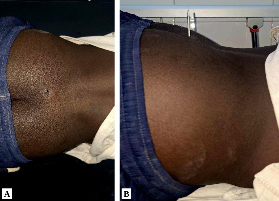
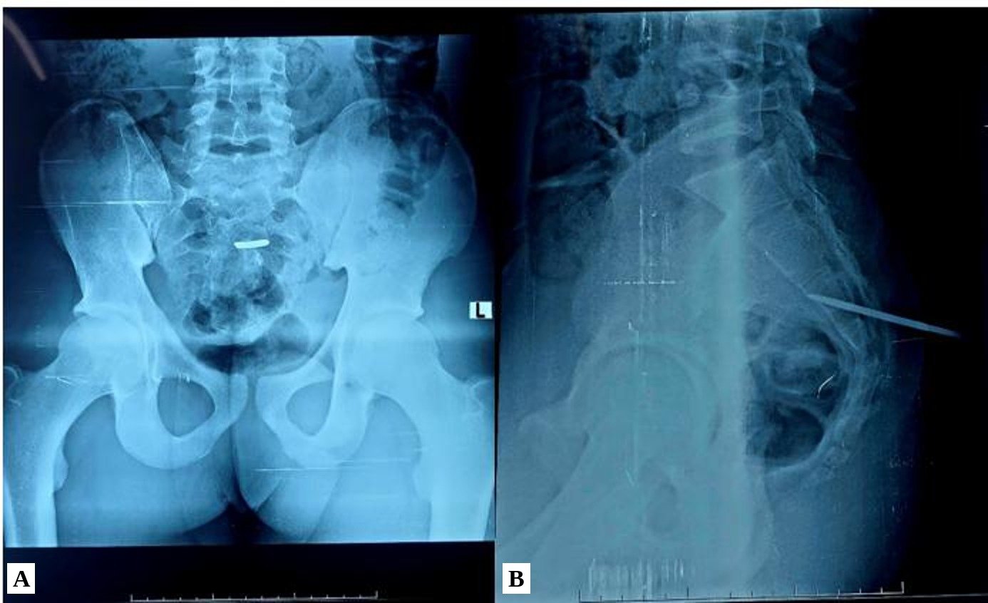
We examined him approximately 11 hours after the injury. There were no neurological signs, no CSF leakage, no sphincter disturbance and no signs of intraperitoneal organ injury. He was classified as ASIA E. Local examination revealed a metal shaft located 1 cm to the right of the midline and 3 cm above the intergluteal cleft, firmly fixed in the deep tissues, with healthy surrounding skin that was tender on palpation. Inspection of the shaft showed that it was the blade of a reversible screwdriver whose handle was missing (Figure 1); the handle had probably come off after the impact. Anteroposterior and lateral radiographs of the lumbosacral spine showed that the shaft crossed the sacral canal and the body of S3 without breaching its anterior wall (Figure 2). There were no signs of hollow viscus perforation and no haematoma along the track. No further imaging could be performed, and we decided to carry out direct extraction in the treatment room, with the surgical and anaesthesia teams on standby to proceed to exploration in the operating theatre if any complication occurred.
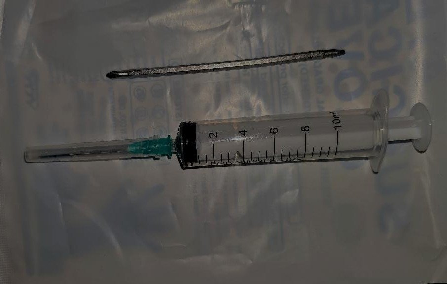
The patient received a dose of antibiotic prophylaxis (amoxicillin–clavulanic acid 2 g IV) and a tetanus booster. The skin was prepared with povidone-iodine, and local anaesthesia was performed around the entry wound with 1% lidocaine (Xylocaine). The shaft was removed with a gouge forceps, which allowed it to be gripped firmly and extracted by traction, taking care to follow the same angle as the entry track seen on the lateral radiograph (Figure 3). The entry wound was sutured, and the patient was discharged home the following day. He was reviewed 15 days, 1 month and 2 months after the injury, with a normal clinical examination and good wound healing.
Non-ballistic penetrating injuries of the spine are rare at all levels, and the available data come mainly from case reports rather than large hospital series [25]. The largest published series come from South Africa [7, 18]. The most recent is that of Seroto et al. in 2021, which included 96 patients, none with a sacral injury [24]. Elsewhere, the largest series are those of Heary et al. and Simpson et al. in the United States and Thakur et al. in India, each with fewer than 5 cases per year [10, 26, 28].
This patient is the first case seen in our department over a 5-year period, and lumbosacral injuries such as this one are less frequent than cervical and thoracic injuries [23]. More specifically, sacral injuries often result from perineal trauma during a fall onto a sharp object, rather than from a direct blow to the sacral region (Table I) [12, 32]. In assaults, the cervical and thoracic regions are targeted more often than the lumbosacral region [21, 28].
The young age and male sex of our patient are consistent with published cases [7, 17, 18, 28].
A wide variety of objects have been described as causes of penetrating spinal injuries, ranging from genuine bladed weapons to glass shards and wood fragments [2, 16, 17, 28]. Assaults are the most common circumstance; the wounding agent may remain intact at the point of impact, be withdrawn by the assailant, or break during an attempt at removal, leaving a foreign body more or less buried and fixed in the deep tissues. This was the case in our patient, in whom the screwdriver handle probably came off after the impact, as the assailant pulled his hand away.
Diagnosis is straightforward when the patient presents with the wounding agent still visible at the point of impact, or when plain radiographs guided by the history reveal it once it is no longer visible on examination. This, however, applies only to radiopaque foreign bodies (metal objects such as knife blades, machetes or chisels). When the foreign body is radiolucent (wood fragment, toothpick, chopstick, etc.), it may be missed on plain radiographs. It may also be overlooked on computed tomography (CT), where its hypodense appearance may suggest air artefacts and mislead the diagnosis [9]. In some cases, diagnosis has therefore been delayed by several weeks or even years after the initial injury [11, 13]. When a foreign body is suspected, close follow-up looking for signs such as progressive compressive myelopathy or suppuration at the entry site will help to establish the diagnosis [9]. Magnetic resonance imaging (MRI) may have a role in such cases, but not as a first-line investigation, because a foreign body that is incompatible with the magnetic field could move and cause damage that was not initially present.
Extraction of a foreign body fixed in the spine can be difficult and may cause neurological sequelae. Extraction procedures are nonetheless not standardised, and the choice between open and closed extraction is made on a case-by-case basis [25, 30]. In our patient, the decision criteria were the absence of CSF leakage and of neurological signs, in particular the absence of any sign of cauda equina root involvement. This may be explained by the fact that the wounding agent crossed the sacral canal at the level of the lower part of the S3 vertebra before heading upwards; Roger et al. showed that the dural sac ends between the middle third of S1 and the lower third of S2 in black African subjects [20]. The absence of a haematoma along the track led us to presume that there was no vascular injury.
Taken together, these findings led us to conclude that further surgical exploration was not warranted, in agreement with the largest series [14, 18]. Moreover, direct removal had already been described by Villarreal-García et al. in Mexico in 2018, in a patient with characteristics similar to ours whose course was uncomplicated, and earlier by Sakar et al. in Turkey in 2016 [22, 29]. This confirms that, in the absence of neurological signs and CSF leakage, direct removal of the foreign body is possible and avoids the complications specific to surgical exploration (dural tear, infection, pain due to muscle detachment, delayed wound healing, prolonged hospital stay, etc.). Antibiotic and tetanus prophylaxis help to prevent delayed infectious complications [31].
Although the history and physical examination allowed us to presume the absence of neurological and associated injuries, plain radiography is insufficient. Contrast-enhanced CT, the reference investigation in this setting to rule out hollow viscus, vascular or urinary tract injury, could not be performed in our patient, partly because the scanner in our hospital was unavailable for maintenance, and partly because of financial constraints related to the patient's socioeconomic situation, which prevented his transfer to another facility for further imaging.
Table I: Published case reports of penetrating injury to the sacrum
| Authors | Number | Location | Wounding agent | Sex | Age (years) | Neurological signs |
|---|---|---|---|---|---|---|
| Gul et al. (2010) [9] | 1 | S1–S2 | Wood | M | 23 | S1–S3 hypoaesthesia |
| Anderson et al. (2010) [1] | 1 | S3 | Glass | M | 6 | Low back pain, dural laceration |
| Brembilla et al. (2015) [3] | 1 | S1 | Pencil | M | 42 | CSF leak |
| Piqueras et al. (2006) [19] | 1 | L5–S1 | Pencil | M | 10 | Cauda equina syndrome |
| Stern et al. (2011) [27] | 1 | S3 | Nail gun | M | 21 | None |
CSF: cerebrospinal fluid; M: male.
Penetrating injuries of the sacrum are uncommon in the literature, and their management is not standardised. It should be guided by careful clinical assessment. Where necessary, and depending on the history, imaging will be required to rule out residual foreign bodies and associated injuries. Removal without surgical exploration therefore remains an option in the sacral region in the absence of neurological signs, CSF leakage, local infection or a foreign body buried beneath the skin, provided that a team is informed and ready to respond to any complication.
We requested and obtained the patient's free and informed consent for the publication of his data. We thank the patient for his consent.
Conflict of interest: None.
*Correspondence: Claude Berny DIBANGOU MAVOUNGOU
Cbmav63@gmail.com
Available online: November 9, 2026
1 Department of Neurosurgery, Idrissa POUYE General Hospital, Dakar, Senegal
2 Faculty of Medicine and Odonto-Stomatology, Dakar, Senegal
3 Department of Neurosurgery, Ziguinchor Regional Hospital, Senegal
© Journal of African Clinical Cases and Reviews 2026
1. Anderson ER, Grant HI, Weissman M. Penetrating Glass Injury to the Sacral Spine. Clinical Medicine & Research. 2010;8(2):114‑115.
2. Baghai P, Sheptak PE. Penetrating spinal injury by a glass fragment: case report and review. Neurosurgery. 1982;11(3):419‑422.
3. Brembilla C, Lanterna LA, Gritti P, Costi E, Dorelli G, Moretti E, et al. Disabling Orthostatic Headache after Penetrating Stonemason Pencil Injury to the Sacral Region. Case Rep Emerg Med. 2015;2015:623405. https://doi.org/10.1155/2015/623405.
4. Burney RE, Maio RF, Maynard F, Karunas R. Incidence, characteristics, and outcome of spinal cord injury at trauma centers in North America. Arch Surg. 1993;128(5):596‑599.
5. Cybulski GR, Stone JL, Kant R. Outcome of laminectomy for civilian gunshot injuries of the terminal spinal cord and cauda equina: review of 88 cases. Neurosurgery. 1989;24(3):392‑397.
6. Daban JL, Peigne V, Boddaert G, Ondo RO, Paul S. TRAUMATISME PÉNÉTRANT ET BALISTIQUE. (SFAR). 2012;
7. Enicker B, Gonya S, Hardcastle TC. Spinal stab injury with retained knife blades: 51 Consecutive patients managed at a regional referral unit. Injury. 2015;46(9):1726‑1733.
8. Gueye EM, Sakho Y, Badiane SB, Ba MC, Diene MS, Diop AA, et al. [Spinal cord injuries in Senegal: 16 cases]. Dakar Med. 1998;43(2):238‑242.
9. Gul S, Dusak A, Songur M, Kalayci M, Acikgoz B. Penetrating Spinal Injury With a Wooden Fragment: A Case Report and Review of the Literature. Spine. 2010;35(25):E1534‑E1536.
10. Heary RF, Vaccaro AR, Mesa JJ, Balderston RA. Thoracolumbar infections in penetrating injuries to the spine. Orthop Clin North Am. 1996;27(1):69‑81.
11. Jones FD, Woosley RE. Delayed myelopathy secondary to retained intraspinal metallic fragment. Case report. J Neurosurg. 1981;55(6):979‑982.
12. Ko J-H, Chong S-W, Nguyen V-L, Ham D-H. Anterior approach for a stab wound with penetrating rebar injury causing incomplete cauda equina syndrome in lumbosacral spine lesion: a case report and literature review. Eur Spine J. 2024;33(7):2870‑2877.
13. Kulkarni AV, Bhandari M, Stiver S, Reddy K. Delayed presentation of spinal stab wound: case report and review of the literature. J Emerg Med. 2000;18(2):209-213. https://doi.org/10.1016/S0736-4679(99)00196-1.
14. Lipschitz R, Block J. Stab wounds of the spinal cord. Lancet. 1962;2(7248):169‑172.
15. Little JW, DeLisa JA. Cauda equina injury: late motor recovery. Arch Phys Med Rehabil. 1986;67(1):45‑47.
16. Opel DJ, Lundin DA, Stevenson KL, Klein EJ. Glass foreign body in the spinal canal of a child: case report and review of the literature. Pediatr Emerg Care. 2004;20(7):468‑472.
17. Pal D, Timothy J, Marks P. Penetrating spinal injury with wooden fragments causing cauda equina syndrome: case report and literature review. Eur Spine J. 2006;15(S5):574‑577.
18. Peacock WJ, Shrosbree RD, Key AG. A review of 450 stabwounds of the spinal cord. S Afr Med J. 1977;51(26):961‑964.
19. Piqueras C, Martínez-Lage JF, Almagro MJ, Ros De San Pedro J, Torres Tortosa P, Herrera A. Cauda equina-penetrating injury in a child. Case report. J Neurosurg. 2006;104(4 Suppl):279‑281.
20. Roger MTJ, Samuel NA, Cathy MZ. Anatomy/Anatomie Terminaison Du Cône Médullaire, Du Sac Dural Et Profondeur Du Canal Vertébral Chez Le Noir Africain Conus Medullaris Position, Dural Sac Level and Vertebral Canal Depth on Black African Subjects. African Journal of Neurological Sciences. 2012;31(2):65.
21. Saeidiborojeni HR, Moradinazar M, Saeidiborojeni S, Ahmadi A. A survey on spinal cord injuries resulting from stabbings: a case series study of 12 years' experience. J Inj Violence Res. 2013;5(1):70‑74.
22. Sakar M, Dogrul R, Niftaliyev S, Bayri Y, Dagcınar A. Direct withdrawal of a knife lodged in the thoracic spinal canal in a patient with normal neurologic examination: is it safe? Spinal Cord Ser Cases. 2016;2:16009.
23. Scheiderer B, Mild K, Gebhard F, Scola A. Penetrierende Stichverletzung des lumbalen Rückenmarks bei einem Kind. Unfallchirurg. 2016;119(3):251‑254.
24. Seroto PM, Harrington BM, Lombard C, Vlok AJ. The role of tertiary neurosurgical intervention in non-missile penetrating injuries of the spine. Eur Spine J. 2021;30(6):1397‑1401.
25. Shahlaie K, Chang DJ, Anderson JT. Nonmissile penetrating spinal injury. Case report and review of the literature. J Neurosurg Spine. 2006;4(5):400‑408.
26. Simpson RK, Venger BH, Narayan RK. Treatment of acute penetrating injuries of the spine: a retrospective analysis. J Trauma. 1989;29(1):42‑46.
27. Stern LC, Moore TA. Nail gun injury to the sacrum: case report and review of the literature. Spine (Phila Pa 1976). 2011;36:E1778-E1780. https://doi.org/10.1097/BRS.0b013e318226771f.
28. Thakur RC, Khosla VK, Kak VK. Non-missile penetrating injuries of the spine. Acta Neurochir (Wien). 1991;113(3‑4):144‑148.
29. Villarreal-García FI, Reyes-Fernández PM, Martínez-Gutiérrez OA, Peña-Martínez VM, Morales-Ávalos R. Direct withdrawal of a knife in the lumbar spinal canal in a patient without neurological deficit: case report and review of the literature. Spinal Cord Ser Cases. 2018;4:48.
30. Yamaguchi S, Eguchi K, Takeda M, Hidaka T, Shrestha P, Kurisu K. Penetrating Injury of the Upper Cervical Spine by a Chopstick -Case Report-. Neurol Med Chir (Tokyo). 2007;47(7):328‑330.
31. Yilmaz N, Kiymaz N, Mumcu C, Demir I. Penetrating spinal injury: reports of two cases. Ulus Travma Acil Cerrahi Derg. 2009;15(1):91‑94.
32. Zhou Z, Song Y, Cai Q, Li T, Liu H. Penetrating Injury of Rectum and Vertebral Body by Steel Bar Causing Cauda Equina Syndrome. Spine. 2011;36(12):E803‑E807.
How to cite this article: Dibangou CBM, Ezabwanga RJM, Ndiaye AD, Sambou NM. Penetrating trauma to the sacrum: a case report and review of the literature. Jaccr Case Reports 2026.
